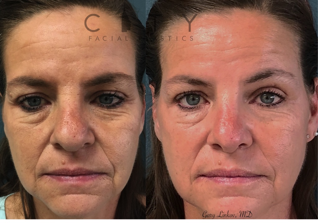
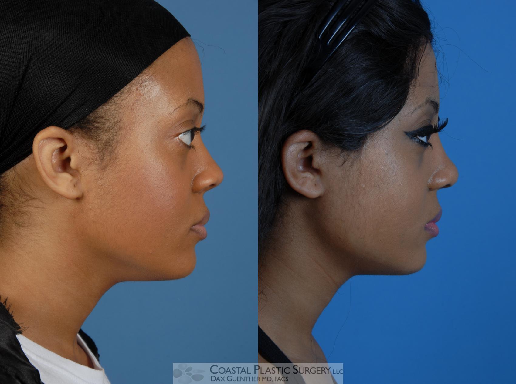
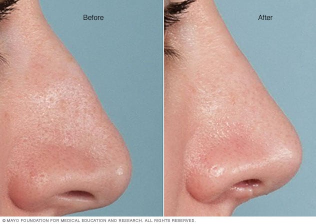
Facts About Rhinoplasty Surgery Austin Revealed
Table of ContentsAustin Rhinoplasty Surgery Things To Know Before You Get ThisNot known Incorrect Statements About Rhinoplasty Surgery Austin Tx Rhinoplasty Surgery Austin Tx - The Facts
Lower third area the skin of the lower nose is as thicker and less mobile, because it has more sebaceous glands, particularly at the nasal pointer. Subcutaneous fat layer is very thin. Nasal lining At the vestibule, the human nose is lined with a mucous membrane of squamous epithelium, which tissue then shifts to become columnar respiratory epithelium, a pseudo-stratified, ciliated (lash-like) tissue with abundant seromucous glands, which maintains the nasal moisture and protects the respiratory system from bacteriologic infection and foreign items.
the elevator muscle group that includes the procerus muscle and the levator labii superioris alaeque nasi muscle. the depressor muscle group which consists of the alar nasalis muscle and the depressor septi nasi muscle. the compressor muscle group which consists of the transverse nasalis muscle. the dilator muscle group which consists of the dilator naris muscle that expands the nostrils; it is in 2 parts: (i) the dilator nasi anterior muscle, and (ii) the dilator nasi posterior muscle.
To prepare, map, and carry out the surgical correction of a nasal problem or deformity, the structure of the external nose is divided into nine (9) visual nasal subunits, and 6 (6) visual nasal sectors, which provide the cosmetic surgeon with the procedures for determining the size, degree, and topographic place of the nasal problem or deformity.
Hence, if more than half of an aesthetic subunit is lost (harmed, malfunctioning, destroyed) the surgeon changes the whole aesthetic segment, generally with a regional tissue graft, harvested from either the face or the head, or with a tissue graft collected from somewhere else on the patient's body. Like the face, the human nose is well vascularized with arteries and veins, and hence supplied with plentiful blood.
The external nose is supplied with blood by the facial artery, which ends up being the angular artery that courses over the superomedial aspect of the nose. The sellar area (sella turcica, "Turkish chair") and the dorsal area of the nose are provided with blood by branches of the internal maxillary artery (infraorbital artery) and the ophthalmic arteries that additional info obtain from the internal typical carotid artery system.
The Greatest Guide To Nose Job Austin
The nasal septum also is supplied with blood by the sphenopalatine artery, and by the anterior and posterior ethmoid arteries, with the additional circulatory contributions of the superior labial artery and of the greater palatine artery - rhinoplasty surgery austin. These three (3) vascular supplies to the internal nose assemble in the Kiesselbach plexus (the Little area), which is an area in the anteroinferior-third of the nasal septum, (in front and listed below).
The nasal veins are biologically considerable, because they have no vessel-valves, and due to the fact that of their direct, circulatory interaction to the spacious sinus, that makes possible the prospective intracranial dispersing of a bacterial infection of the nose. For this reason, due to the fact that of such a plentiful nasal blood supply, tobacco smoking cigarettes does therapeutically compromise post-operative healing.
Nasal innervation: Cranial nerve V, the trigeminal nerve (nervus trigeminis) gives feeling to the nose, the face, and the upper jaw (maxilla) - rhinoplasty surgery austin. The experiences registered by the human nose derive from the very first two (2) branches of cranial nerve V, the trigeminal nerve. The nerve listings show the respective innervation (sensory circulation) of the trigeminal nerve branches within the nose, the face, and the upper jaw (maxilla).
The indicated nerve serves the named anatomic facial official source and nasal areas Lacrimal nerve conveys sensation to the skin locations of the lateral orbital (eye socket) region, except for the lacrimal gland. Frontal nerve communicates experience to the skin areas of the forehead and the scalp. Supraorbital nerve conveys experience to the skin areas of the eyelids, the forehead, and the scalp.
Nasociliary nerve communicates feeling to the skin location of the nose, and the mucous membrane of the anterior (front) nasal cavity. Anterior ethmoid nerve conveys sensation in the anterior (front) half of the nasal cavity: (a) the internal areas of the ethmoid sinus and the frontal sinus; and (b) the external locations, from the nasal suggestion to the rhinion: the anterior pointer of the terminal end of the nasal-bone suture.
Infratrochlear nerve conveys experience to the median area of the eyelids, the palpebral conjunctiva, the nasion (nasolabial junction), and the bony dorsum. Nasal anatomy: The shell-form turbinates (conchae nasales). Nasal anatomy: The septum nasi bones and cartilages. The supply of parasympathetic nerves to the face and the upper jaw (maxilla) stems from the greater shallow petrosal (GSP) branch of cranial nerve VII, the facial nerve.
Rhinoplasty Austin Can Be Fun For Anyone
The floor of the nose comprises the premaxilla bone and the palatine bone, the roof of the mouth. The nasal septum is composed of the quadrangular cartilage, the vomer bone (the perpendicular plate of the ethmoid bone), elements of the premaxilla, and the palatine bones. Each lateral nasal wall includes find more info 3 pairs of turbinates (nasal conchae), which are little, thin, shell-form bones: (i) the superior concha, (ii) the middle concha, and (iii) the inferior concha, which are the bony framework of the turbinates.